image
(CT) imaging of the left breast and axilla
0 views
Mar 16, 2026
  • Diagnosis Title: Invasive ductal carcinoma
  • Disease Specialty: Surgery
  • Ease of Diagnosis: Difficult
  • Certainty: Certain
  • Ethnicity: Asian
  • Segment: Adult Male
  • Image Quality: High
  • Clinical Examination: The patient appeared to be generally well with a normal distribution of male body hair, no gynecomastia, and body mass index (BMI) of 36.9 kg/m2. Physical examination of the left breast and axilla showed a hard, erythematous, ulcerating, mass measuring approximately 9×5 cm in the subareolar region. His left nipple was distorted by the mass with no discharge. His body temperature was normal. Examination of the ipsilateral axilla showed an enlarged mobile lymph node. The right breast and axilla were normal on examination.
  • Patient Age: 59 year
  • Patient Gender: Male
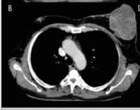
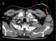
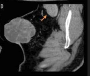
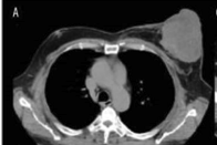
  • A 59-year-old man presented with a large left breast mass with enlarged axillary lymph nodes. The patient had ignored the mass and declined all diagnostic procedures. After modifying the diagnostic workup and involving a psychiatrist, the patient agreed to undergo a modified radical mastectomy. Histopathology showed a high-grade invasive ductal carcinoma with lymph node metastasis. The breast cancer was triple-positive for human epidermal growth factor receptor 2 (HER2), estrogen receptor (ER), and progesterone receptor (PR). Adjuvant treatment included herceptin, tamoxifen, and radiation therapy.